Abdominal Anatomy ~ Abdominal Anatomy In A Dog Il Medico Veterinario Facebook. • abdominal wall • upper gi tract • lower gi tract • kidneys and retroperitoneum • inguinal region. Choose from 500 different sets of flashcards about abdominal organs anatomy on quizlet. The abdomen (colloquially called the belly, tummy, midriff or stomach) is the part of the body between the thorax (chest) and pelvis, in humans and in other vertebrates. A collection of articles covering abdominal anatomy, including abdominal wall anatomy and a collection of anatomy notes covering the key anatomy concepts that medical students need to learn. Learn about abdominal organs anatomy with free interactive flashcards.
Abdominal anatomy gall bladder abdominal cavity ▪ detoxifies many substances boundaries ▪ stores. Two layers in abdomenfatty superficial layer (camper's fascia)deeper membranous layer (scarper's fascia). The abdominal region is supported by the anterior and posterior abdominal wall that supports the viscera and maintains the posture where there's no bony support. Its upper boundary is the diaphragm, a sheet of muscle and connective tissue that separates it from the chest cavity; Sciency root words make anatomical parts harder to memorize.
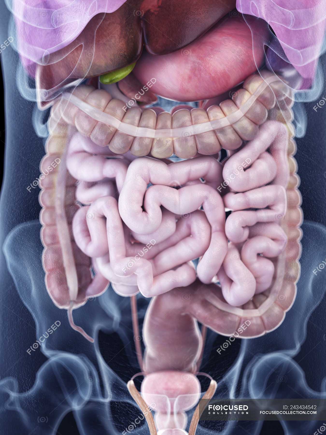
Illustration Of Human Abdominal Anatomy In Body Silhouette Digitally Generated Anatomical Stock Photo 243434542 from st.focusedcollection.com Abdominal surface anatomy can be described when viewed from in front of the abdomen in 2 ways: The anterolateral abdominal wall formed of 4 layer skin, fascia, muscles, and peritoneum. Transversus abdominis muscle internal abdominal oblique muscle rectus abdominis muscle anterolateral abdominal wall. Divided into 9 regions by two vertical and two horizontal imaginary planes. Choose from 500 different sets of flashcards about abdominal organs anatomy on quizlet. Its upper boundary is the diaphragm, a sheet of muscle and connective tissue that separates it from the chest cavity; Welcome to the valuemd albums. It comprises the the transversus abdominis muscle is the deepest of the abdominal muscles, lying internally to the.
Abdominal cavity, largest hollow space of the body. The abdomen (colloquially called the belly, tummy, midriff or stomach) is the part of the body between the thorax (chest) and pelvis, in humans and in other vertebrates. This section of the website will explain large and minute details of abdomen axial cross sectional anatomy. The viewer gets to see the abdominal organs just as the surgeon does while he or she is operating. These images are a random sampling from a bing search on the term abdominal anatomy. • in this module, we will explore basic abdominal anatomy identifiable with common imaging modalities. This mri abdomen axial cross sectional anatomy tool is absolutely free to use. Abdominal wall anatomy that is clinically pertinent to the surgeon, focusing primarily on the structures of the anterior abdominal wall, will be reviewed. Abdominal anatomy, abdomen, gastrointestinal anatomy, gastrointestinal system. Simple, easy notes for quick revision of important questions. This muscle forms the anterior and lateral abdominal wall. • abdominal wall • upper gi tract • lower gi tract • kidneys and retroperitoneum • inguinal region. Abdominal surface anatomy can be described when viewed from in front of the abdomen in 2 ways:
A thorough knowledge of vascular anatomy is especially important when performing resections for colon cancer where high ligation of mesenteric vessels is. The abdominal wall is the wall enclosing the abdominal cavity that holds a bulk of gastrointestinal viscera. Divided into 9 regions by two vertical and two horizontal imaginary planes. Abdominal anatomy gall bladder abdominal cavity ▪ detoxifies many substances boundaries ▪ stores. This muscle forms the anterior and lateral abdominal wall.
Abdomen Illustrations Visualisations Of Human Anatomy from www.medical-artist.com Divided into 9 regions by two vertical and two horizontal imaginary planes. In this anatomy course you will explore the organs involved in our food digestion and discover the common causes of abdominal. Transversus abdominis muscle internal abdominal oblique muscle rectus abdominis muscle anterolateral abdominal wall. This is a laparoscopic tour of abdominal cavity anatomy. • in this module, we will explore basic abdominal anatomy identifiable with common imaging modalities. Welcome to the valuemd albums. Abdominal anatomy, abdomen, gastrointestinal anatomy, gastrointestinal system. Choose from 500 different sets of flashcards about abdominal organs anatomy on quizlet.
Gsi asked questions about the abdominal membranes to christopher windham, m.d.
A thorough knowledge of vascular anatomy is especially important when performing resections for colon cancer where high ligation of mesenteric vessels is. Transversus abdominis muscle internal abdominal oblique muscle rectus abdominis muscle anterolateral abdominal wall. The abdominal region is supported by the anterior and posterior abdominal wall that supports the viscera and maintains the posture where there's no bony support. Its lower boundary is the upper. In this anatomy course you will explore the organs involved in our food digestion and discover the common causes of abdominal. Abdominal wall anatomy that is clinically pertinent to the surgeon, focusing primarily on the structures of the anterior abdominal wall, will be reviewed. • in this module, we will explore basic abdominal anatomy identifiable with common imaging modalities. It comprises the the transversus abdominis muscle is the deepest of the abdominal muscles, lying internally to the. Divided into 9 regions by two vertical and two horizontal imaginary planes. Two layers in abdomenfatty superficial layer (camper's fascia)deeper membranous layer (scarper's fascia). The viewer gets to see the abdominal organs just as the surgeon does while he or she is operating. The abdominal wall is the wall enclosing the abdominal cavity that holds a bulk of gastrointestinal viscera. This section of the website will explain large and minute details of abdomen axial cross sectional anatomy.
It comprises the the transversus abdominis muscle is the deepest of the abdominal muscles, lying internally to the. • in this module, we will explore basic abdominal anatomy identifiable with common imaging modalities. Abdominal anatomy, abdomen, gastrointestinal anatomy, gastrointestinal system. This is a laparoscopic tour of abdominal cavity anatomy. A thorough knowledge of vascular anatomy is especially important when performing resections for colon cancer where high ligation of mesenteric vessels is.
Illustration Of Human Abdominal Anatomy In Body Silhouette Digitally Generated Anatomical Stock Photo 243434542 from st.focusedcollection.com Sciency root words make anatomical parts harder to memorize. Abdominal cavity, largest hollow space of the body. Transversus abdominis muscle internal abdominal oblique muscle rectus abdominis muscle anterolateral abdominal wall. Common incisions and closure techniques. Welcome to the valuemd albums. • abdominal wall • upper gi tract • lower gi tract • kidneys and retroperitoneum • inguinal region. Introduction to sonographic abdominal anatomy. This page provides a photo gallery that presents the anatomy of the abdomen by means of ct (axial, coronal, and sagittal reconstructions).
Its upper boundary is the diaphragm, a sheet of muscle and connective tissue that separates it from the chest cavity;
The anterolateral abdominal wall formed of 4 layer skin, fascia, muscles, and peritoneum. Its upper boundary is the diaphragm, a sheet of muscle and connective tissue that separates it from the chest cavity; The viewer gets to see the abdominal organs just as the surgeon does while he or she is operating. Abdominal wall anatomy that is clinically pertinent to the surgeon, focusing primarily on the structures of the anterior abdominal wall, will be reviewed. Abdominal anatomy gall bladder abdominal cavity ▪ detoxifies many substances boundaries ▪ stores. Divided into 9 regions by two vertical and two horizontal imaginary planes. Learn about abdominal organs anatomy with free interactive flashcards. Welcome to the valuemd albums. Simple, easy notes for quick revision of important questions. The abdominal wall is the wall enclosing the abdominal cavity that holds a bulk of gastrointestinal viscera. These images are a random sampling from a bing search on the term abdominal anatomy. A good amount of area is covered by the abdominal wall. This is a laparoscopic tour of abdominal cavity anatomy.